A coloured scanning electron micrograph (SEM) of the head of a maggot or the larva of a bluebottle fly (Protophormia sp.) with tiny teeth-like fangs extending from its mouth. The maggots of this fly are used medicinally to clean wounds. The maggots are sterilised and placed in the wound, where they feed on dead tissue and leave healthy tissue untouched. Their saliva contains anti- bacterial chemicals which maintain sterility in the area. Maggots are used on ulcers and deep wounds away from organs or body cavities, most often being used to treat diabetic ulcers on the feet.
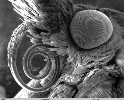
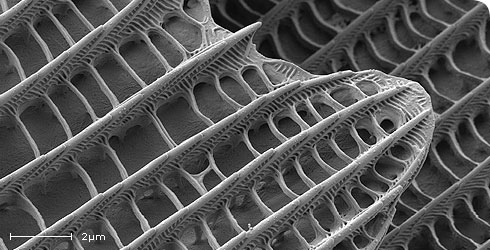
Butterfly Tongue Under The Scanning Electron Microscope
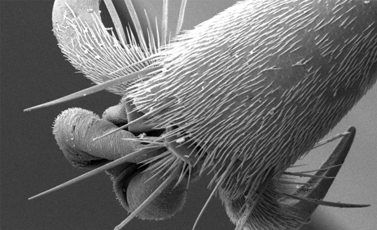
Hornet Leg Under The Scanning Electron Microscope
Triturus Under The Scanning Electron Microscope
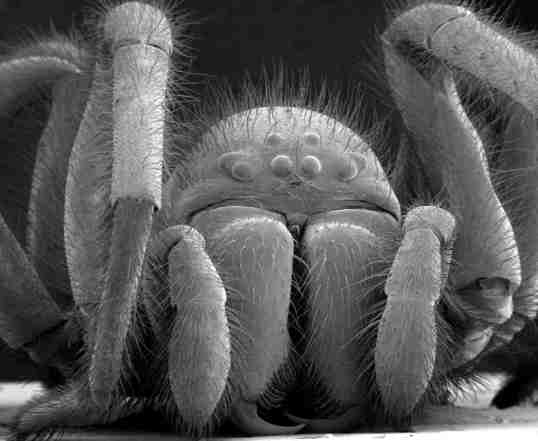
Spider Under The Scanning Electron Microscope
Metastatic Human Prostate Cancer Cell
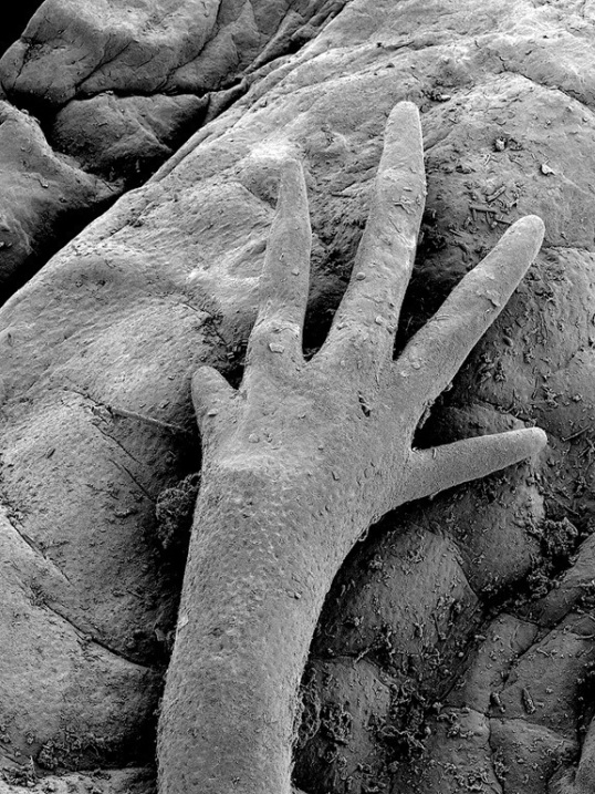
I'm not sure I want to know...
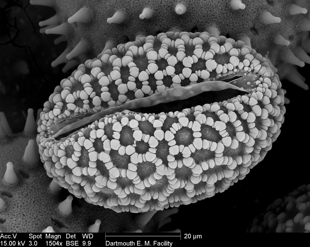
Pollen as seen under a scanning electron microscope.
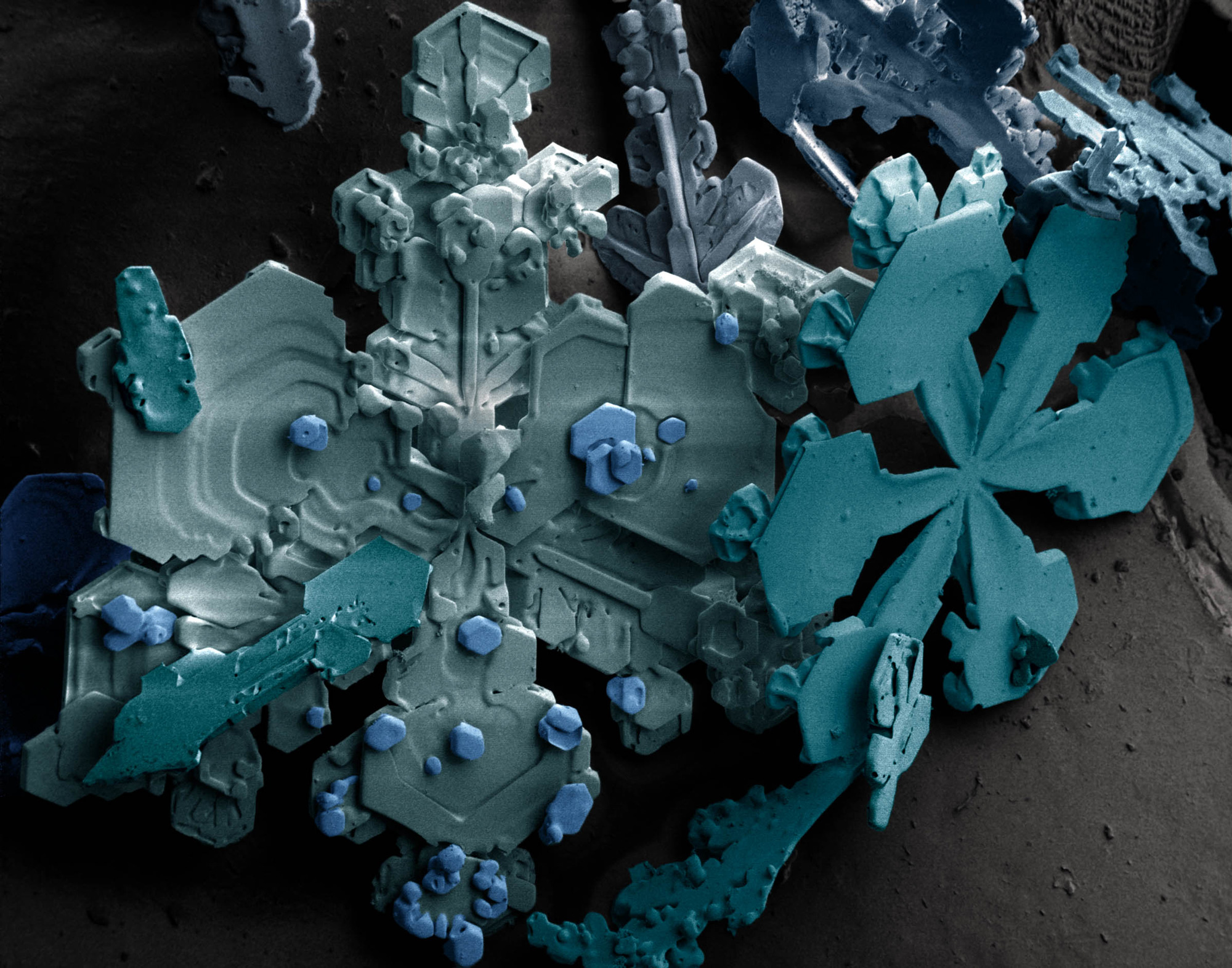
Once snow crystals form in the atmosphere, they grow by absorbing surrounding water droplets. The snowflakes we end up seeing on the ground are an accumulation of these ice crystals.
A thin layer of gold on top of rubber
Here are two stunningly glorious closeup shots of biochar as seen by an electron microscope.
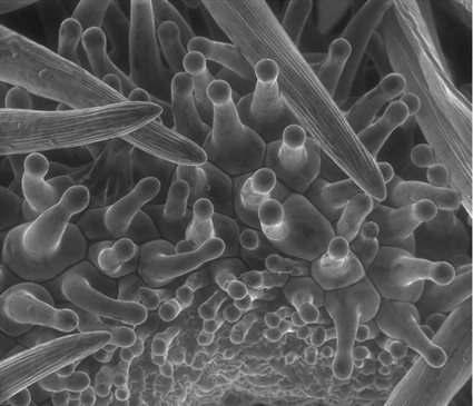
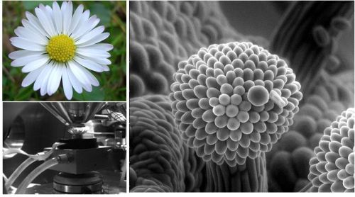
Environmental Scanning Electron Microscopy (ESEM) Image of the surface of a flower.
Fossil of Globigerinelloides Globigerinelloides prairiehillensis (Pessagno, 1967) in edge-on view. Age: approximately 83 million years. This genus had their maximum diversity between 83 and 65 million year.
fungal hyphae of Telephora terrestris and a Nanhermannia sp. mite
Secondary electron images of butterfly scales from Vanessa atalanta. Acquired using the Ultra Plus charge compensation system.